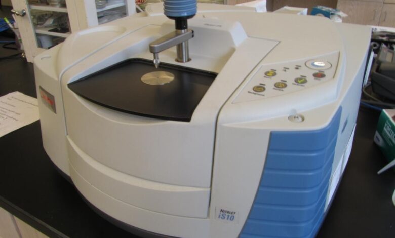
Fourier Transform Infrared (FTIR)Spectroscopy

The rapid evolution of microorganisms has enabled the development of precise techniques for analysis. FTIR spectroscopy methods distinguish a healthy organ from pathological samples. Rapid growth in this field has resulted in more accessible and more impartial diagnoses in the biomedical field.
Applications Of FTIR In Cancer Diagnosis
Fourier Transformed spectroscopy techniques distinguish between cancerous and normal blood tissue. Biomedicine IR-based methods have shown tremendous potential and are now a reality due to the large volume of data accumulated from trials, clinical studies, and developments. Spectroscopy has been applied in recent developments to distinguish cancerous cells from healthy ones. Various tests indicate that the bands of lipids, proteins, nucleic acids, and carbohydrates from cancerous samples are different from the healthy ones. The wavelength band for a healthy tissue is strong and sharp.
Label-free FTIR is used for the early detection of cancer. Studies conducted among various patients’ plasma samples have enabled the development of patterns to detect non-small cell lung carcinoma(NSCLC). An automatic FTIR analysis lab spectroscopic system coupled with pattern recognition algorithms including linear discriminant analysis and random forest clarification is used to study samples.
Recent developments using ATR-FTIR spectroscopy have enabled skin cancer diagnosis and differentiated cancer cell growth potential. Evaluation of the altercations in molecular changes and hydration level using IR spectroscopy has necessitated the identification of different types of cancer.
Applications Of FTIR Spectroscopy Integrated With Lab On A Chip Devices
FTIR spectroscopy has been introduced to modern technologies. Lab-on-chip technology has revolutionized the medical field due to minor equipment for monitoring and diagnosis. The combination of IR radiation and this technology has enabled the integration of microfluidic devices for sugar identification. Efforts have been made to create a low-cost IR Live chip for real-time infrared imaging of living tissues. IR imaging has enabled the distinguishing of cellular organelles and has necessitated the identification of the chemical composition of different tissues at a functional group level.
Recent advances have enabled the development of rapid ATR-FTIR instrumentation. Microvision labs Edx analysis and monitoring of solute concentrations allows the interface of commercially available gadgets with customized microfluidic devices. Technologies like these enable the on-chip identification of chemical compounds, studies of reaction kinetics, and measurement of their concentration in solutions.
Plant Cell Spectromicroscopy
Soft Xray Spectromicroscopy
The lentil stem cells are visible with the help of a light microscope. Internal structures or compositional differences are, however, not detailed. X-ray images show various regions such as cell walls, individual cells, cell components, and the middle lamella. Different polymers have varying absorption intensities, and it is critical to have a method to determine the spatial distribution of lignin. A detailed analysis of the lentil stem section uses spectra of cellulose and lignin, to select and map the number of biopolymers present.
FTIR Spectromicroscopy
Optical microscopic images of the entire lentil stem show the various cell components. Scanned areas show the spatial correlation and distribution of lignin and cellulose. However, this approach does not differentiate individual cells and cell components. Extracted spectra from the regions on the stem indicate that cellulose and lignin are present in highlighted locations.
Relative Merits Of Soft Xray And FTIR Spectromicroscopy Techniques
Infrared methods have less radiation due to minimal intense beams. Keep live-cell samples after data collection. Radiation damage alters the spectral details of biopolymers. Plant samples have complex molecular environments due to many different biopolymers. Complex mixtures spectra are dominated by the absorption of a higher concentration polymer. Antibodies and fluorescent probes have been utilized to identify specific subcellular components and proteins. The use of X-ray excited fluorescent probes with high resolution has enabled the limitation of sensitivity of the biological samples.
Various spectroscopic techniques have been developed, and FTIR can distinguish pathological from healthy samples. Numerous works have been designed to integrate FTIR with ATR techniques to simplify the spectral result.


